
Exploring X-Rays: Discovery, Benefits, Side Effects & Advances
Delve into the fascinating world of X-rays, from their discovery to the benefits, side effects, and modern advances in technology. Learn more about the impact of X-rays on medical diagnostics and treatment.
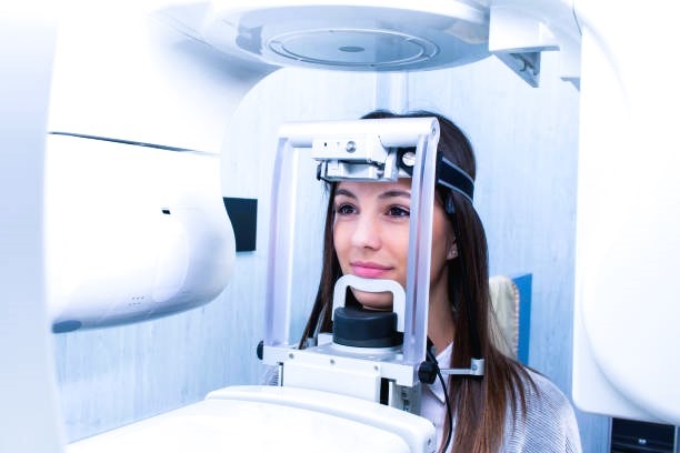
X-rays represent a remarkable form of electromagnetic radiation, characterized by wavelengths shorter than ultraviolet light but longer than gamma rays. Discovered by Wilhelm Conrad Röntgen in 1895, X-rays revolutionized multiple fields but found preeminent utility in the realm of medical imaging. By leveraging their unique properties, X-rays enable physicians to peer inside the human body without invasive procedures, providing crucial insights into patient health.
At the core of X-ray function lies the principle of ionizing radiation. This term refers to the ability of X-rays to ionize atoms and molecules, effectively stripping away electrons and creating charged particles. Such energy transfer allows X-rays to penetrate various materials, a feature that underpins their diagnostic capability. When X-rays pass through the body, denser materials such as bones absorb more radiation, while softer tissues allow more X-rays to pass through. This differential absorption results in detailed contrast images, facilitating the identification and diagnosis of various conditions.
Beyond medical imaging, X-rays are instrumental in several other sectors, including industrial inspection, security, and scientific research. For example, X-ray crystallography has been pivotal in determining the atomic structure of complex molecules, including DNA. Additionally, security systems at airports utilize X-rays to scan luggage and ensure the safety of passengers.
As we delve deeper into the fascinating world of X-rays, we will explore their myriad benefits, potential side effects, and recent advancements that continue to push the boundaries of what this incredible technology can achieve. The profound impact of X-rays on both everyday life and specialized fields underscores their enduring significance and the continual necessity for innovations in this dynamic area of science.
The Discovery of X-Rays
The discovery of X-rays marked a significant milestone in the field of physics and medical science. This revolutionary discovery was made by Wilhelm Conrad Roentgen, a German physicist, in 1895. Roentgen’s work would eventually open new horizons in diagnostic medicine, earning him the first Nobel Prize in Physics in 1901. His curiosity was piqued while experimenting with cathode rays, leading to the unexpected observation of a new type of ray capable of penetrating various substances, which he initially termed "X-rays" due to their unknown nature.
Roentgen's experiments showed that these rays could pass through materials that were opaque to visible light, such as wood, paper, and even human tissue, while being absorbed by denser objects like bones and metal. This penetrating ability of X-rays was first visualized in a striking manner when Roentgen captured an image of his wife’s hand, clearly showing the bones and her wedding ring—this image would become the first-ever medical X-ray.
The implications of Roentgen's discovery were profound and immediate. For the first time in history, it was possible to look inside the human body without making an incision. This groundbreaking method allowed for the detailed, non-invasive examination of bones, paving the way for the development of diagnostic radiology. Roentgen's work not only advanced scientific understanding but also initiated a new era in medicine, where X-rays became indispensable tools for diagnosing fractures, locating foreign objects, and identifying diseases.
Roentgen’s achievement laid the foundation for further research and development in the field of radiography, leading to advancements such as computed tomography (CT) scans and magnetic resonance imaging (MRI). The discovery of X-rays remains a testament to the profound impact of scientific exploration and its potential to transform and save lives.
Benefits of X-Rays
X-rays have revolutionized multiple fields since their discovery, with their most notable impact in medical diagnostics. By providing the ability to observe internal structures without invasive procedures, X-rays have become a crucial tool in modern healthcare. For instance, they are indispensable for detecting fractures, as they can reveal breaks and bone defects that are not visible through a physical examination alone. Additionally, X-rays are used to diagnose and monitor infections, such as pneumonia, by showing areas of lung congestion that signify the presence of the disease.
Beyond detecting physical fractures and infections, X-rays are also crucial in identifying abnormalities within various organs and tissues. For instance, mammography, a specialized X-ray technique, plays a pivotal role in screening for breast cancer by highlighting unusual masses or calcifications. Dental X-rays, another specialized adaptation, allow for precise identification of cavities, root infections, and structural anomalies in the jaw and teeth.
The utility of X-rays extends beyond the realm of healthcare. In the field of security, especially at airports, X-ray scanners have become standard tools for inspecting luggage. These scanners can pinpoint potentially dangerous items by producing detailed images of the contents without having to physically open bags. This enhances security measures while expediting the screening process.
In industry, X-rays serve a significant role in non-destructive testing methods, particularly for evaluating the integrity of materials and constructions. For example, they can be employed to inspect welds within pipelines, detecting any faults or weaknesses that could lead to failures or leaks. This application is critical in maintaining safety standards in sectors such as oil and gas, manufacturing, and civil engineering.
Furthermore, X-rays have widened the scope of research across multiple scientific disciplines. From probing the structure of materials in physics to analyzing archeological artifacts without causing damage, their applications are diverse and far-reaching. Thus, the advent of X-rays has not only enhanced the precision and efficiency of medical diagnostics but has also provided invaluable tools across various industries and research fields, underscoring their multifaceted benefits.
Side Effects and Risks of X-Rays
X-rays, while indispensable in medical diagnostics, come with certain side effects and risks that require careful consideration. Understanding the concept of radiation dose is critical. The radiation dose is the amount of ionizing radiation energy absorbed by the body, measured in units such as Sieverts (Sv) or millisieverts (mSv). The severity of side effects and risks correlates with the level and frequency of exposure, adhering to the principle that higher doses pose greater risks.
One of the primary concerns is the accumulation of radiation over time. Cumulative exposure can lead to long-term health implications. For instance, radiation burns, though rare in diagnostic imaging, can occur if the skin is exposed to high doses in a short period. More commonly, increased cumulative exposure to X-rays has been linked to a slightly elevated risk of developing cancer. Ionizing radiation can cause changes in cell structure and DNA, with prolonged or repeated exposure raising the likelihood of malignant transformations.
To mitigate these risks, several precautions are routinely implemented. Healthcare providers use protective measures such as lead aprons, thyroid shields, and other barriers to shield patients from unnecessary radiation exposure. Additionally, medical professionals are trained to limit the use of X-rays to situations where they are medically justified. Modern imaging technology also aids in minimizing exposure by optimizing radiation doses to the lowest possible levels necessary for accurate diagnostics.
Health authorities, including the World Health Organization (WHO) and various national radiological protection boards, provide guidelines on safe levels of exposure. These guidelines emphasize the importance of the ALARA principle—As Low As Reasonably Achievable—urging the reduction of radiation doses to the minimum necessary to achieve diagnostic benefits. Regular updates to protocols ensure that advancements in imaging technologies and methodologies are integrated to enhance patient safety.
Latest Advances in X-Ray Technology
Recent advancements in X-ray technology have significantly transformed medical imaging, providing clearer images, reducing patient exposure to radiation, and introducing novel diagnostic tools. Enhanced imaging quality is one of the most notable improvements, achieved through the development of high-definition detectors and sophisticated image processing algorithms. These innovations allow healthcare professionals to obtain more accurate and detailed images, aiding in precise diagnoses and better patient outcomes.
Another significant advancement is the reduction of radiation doses. Modern X-ray machines are designed to minimize radiation exposure while maintaining image quality. Techniques such as adaptive exposure control and iterative reconstruction algorithms have been implemented to ensure that the lowest possible dose is used without compromising diagnostic accuracy. This is particularly beneficial for pediatric patients and individuals requiring frequent imaging.
The transition from analog to digital X-rays has been another transformative shift. Digital X-ray systems offer several advantages, including faster image acquisition, easier image storage and retrieval, and improved image manipulation capabilities. The ability to enhance images digitally allows for better visualization of soft tissues and subtle abnormalities that may not be as easily detectable with traditional film-based X-rays.
Computed tomography (CT) scans represent a groundbreaking advancement in X-ray technology. By capturing multiple X-ray images from different angles and reconstructing them into a three-dimensional image, CT scans provide intricate cross-sectional views of the body's internal structures. This capability is invaluable in diagnosing complex conditions, guiding surgical interventions, and monitoring treatment progress.
Dual-energy X-rays are another innovative technology, enabling the differentiation of materials with similar densities. This technique is particularly useful in applications such as identifying bone density variations and detecting certain types of cancer. Portable X-ray machines have also emerged, allowing for on-site imaging in various settings, including remote locations, sports events, and disaster zones. These devices offer high mobility without sacrificing image quality, making X-ray diagnostics more accessible.
Ongoing research and emerging technologies continue to push the boundaries of X-ray technology. For instance, photon-counting detectors promise even higher resolution images and further reductions in radiation exposure. AI-driven image analysis and machine learning algorithms are also being explored to enhance diagnostic accuracy and facilitate automated, real-time image interpretation. These advancements hold the potential to revolutionize X-ray technology, making it an even more powerful tool in medical diagnostics and treatment planning.
Conclusion and Future Outlook
The discovery of X-rays in 1895 by Wilhelm Conrad Roentgen revolutionized the fields of medicine, security, and scientific research. Initially a breakthrough in diagnostic imaging, X-rays have become invaluable for non-invasive internal inspections, enabling earlier and more accurate diagnoses. Moreover, the benefits of X-rays have extended beyond the medical realm; they play a pivotal role in security measures, such as airport baggage scanning, and in various scientific applications including material analysis and art restoration.
Despite their numerous advantages, the exposure to X-rays does come with certain risks, primarily due to ionizing radiation. Side effects, such as tissue damage and increased cancer risks, particularly from repeated or high-dose exposure, necessitate stringent safety protocols. Advances in technology have significantly mitigated these risks through lower radiation doses and improved imaging techniques. As researchers continue to optimize the technology, it is expected that the balance between diagnostic value and patient safety will continually improve.
Looking ahead, the future potential of X-rays seems boundless. Ongoing research and technological enhancements are leading to innovations like 3D imaging, digital radiography, and portable X-ray devices, which promise even greater accuracy and versatility. Additionally, the integration of artificial intelligence in image analysis is set to revolutionize diagnostic practices further by providing faster and more accurate interpretations.
In essence, X-rays remain a critical tool across multiple sectors. Their capacity for deep insight into otherwise inaccessible areas ensures their continued relevance and utility. To leverage these advancements beneficially, staying informed about the latest developments and adopting best practices for safety and efficacy in their use is of paramount importance. The journey of understanding and improving X-rays is far from over, and the future holds vast potential for this remarkable technology.